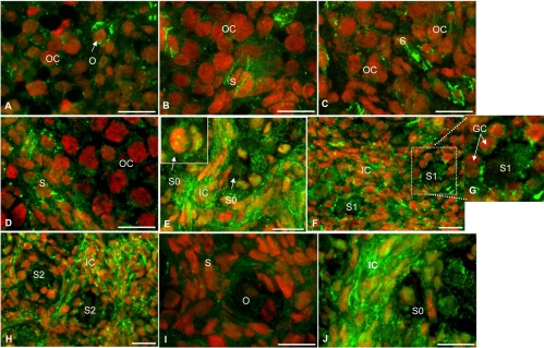
Figure 5.
Immunofluorescence localization of BMPRIB protein in hamster ovary sections during perinatal development and after in vivo exposure to FSH antiserum. For the sake of brevity, images of selected days are furnished to highlight the pattern of developmental expression. E13 and E15 (A and B), P3 and P6 (C and D), P8 (E, insert) show receptor expression in the granulosa cells and oocyte of a primordial follicle. P10 (F and G, insert) show increased BMPRIB expression at the oocyte plasma membrane and granulosa cells interface of an S1 follicle on P10. P15 (H) and ovaries of P8 hamsters (I and J) exposed in utero to an FSH antiserum on E12 and received a single injection of saline (I) or eCG (J) on P1. OC, Oocyte cluster; O, oocyte; S, somatic cells; GC, granulosa cell; S0, primordial follicle; S1, primary follicle; S2, preantral follicles at stage 2; IC, interstitial cells.

