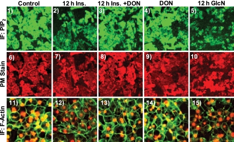
Figure 3.
Hyperinsulinemic and GlcN states induce a loss in PM PIP2 detection and cortical F-actin. Representative immunofluorescent images of PIP2 (panels 1–5) and WGA (panels 6–10) detected in PM sheets treated as described in preceding figures are shown. Phalloidin-stained F-actin and propidium iodide-labeled nuclei in these cells (panels 11–16) are shown. Images are representative from three to five independent experiments.

