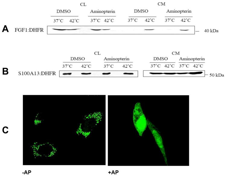
Figure 2. AP does not prevent the secretion of FGF1:DHFR and 6Myc:S100A13:DHFR.
NIH 3T3 cells were transiently transfected with FGF1:DHFR (A) or 6Myc:S100A13:DHFR (B). Transfected cells were pre-incubated for 4 hours with 1 mM AP or with DMSO (solvent control). Then, cells were incubated for 2 hours at 37°C or 42°C, and CL and conditioned media (CM) were collected. FGF1:DHFR and 6Myc:S100A13:DHFR were isolated from CM, resolved by SDS-PAGE and immunoblotted with anti-DHFR antibodies. (C). AP in the concentration of 1 mM abolishes mitochondrial translocation of MTS:GFP:DHFR reporter. NIH 3T3 cells were transfected with MTS:GFP:DHFR. AP was added to the cells 8 h after transfection. Cells were incubated for additional 4 h with or without AP and then fixed. Confocal microscopy (objective X63) demonstrated that incubation with AP results in diffuse cytosolic and nuclear distribution of MTS:GFP:DHFR instead of punctate mitochondrial.

