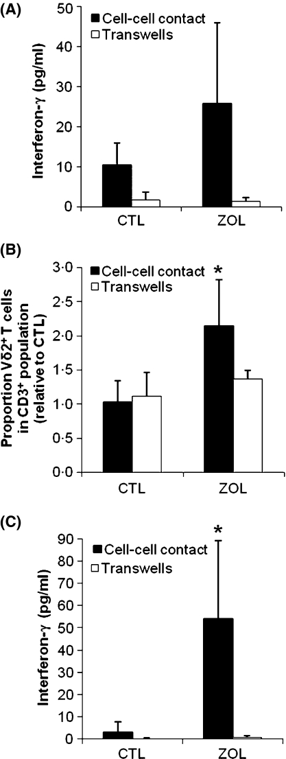
Fig 2.
Activation of Vγ9Vδ2 T cells by ZOL-treated monocytes. Monocytes were purified from human PBMCs by CD14 magnetic bead isolation and pulsed with vehicle or 1 μmol/l ZOL for 2 h. Vehicle- or ZOL-treated monocytes were then co-cultured with monocyte-depleted PBMCs (A, B) or γδ T cells obtained from monocyte-depleted PBMCs using a γδ T cell magnetic bead isolation kit (C). Transwell inserts were used to separate the monocytes from the PBMCs or the enriched γδ T cells. After 72 h, the concentration of IFN-γ in the cell culture medium was determined by enzyme-linked immunosorbent assay (A, C). To determine proliferative responses, the proportion of Vδ2+ T cells in the CD3+ population was determined by immunolabelling and flow cytometric analysis after 7 d of culture and expressed relative to control (B). Data is shown as mean ± standard deviation of 3 independent donors. *P< 0·05 as compared to vehicle-treated control.

