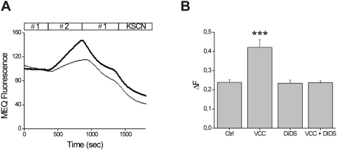
Figure 2. Chloride efflux induced by VCC in Caco-2 cells.
A) Caco-2 cells, loaded with MEQ were incubated with (thick line) or without (thin line) 500 pM VCC. Fluorescence was estimated upon sequential buffer substitution with Cl−- containing medium (# 1) followed by Cl−-free medium (# 2). Substitution of solution (#1) by solution (#2) led to an increase of MEQ fluorescence (dequenching). Subsequent substitution by solution #1 induced an influx of Cl− ions, thus quenching MEQ fluorescence. Values are normalized with respect to the base line. B) Effect of 100 µM DIDS on VCC-induced chloride efflux. ΔF has been calculated as reported in the Material and Methods section. Each histogram represents an average of thirteen independent experiments with a recording of 2–3 cells in each assay. Significance, determined by Student's t test, was compared to non-treated cells (Ctrl); ***, p<0.001.

