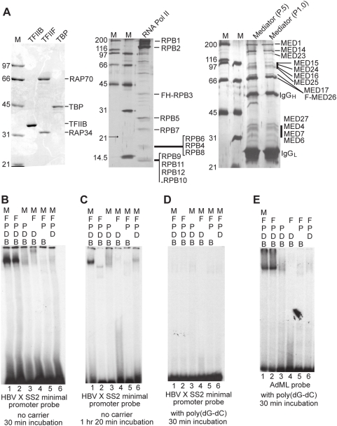
Figure 10. Sequence-nonspecific stable complex formation by TBP, TFIIB, RNA pol II, TFIIF, and mediator on X gene Start site 2 promoter.
(A) SDS-PAGE analyses of purified factors. Purified recombinant TFIIB, TFIIF, and TBP were analyzed by Comassie staining. RNA pol II and mediator were analyzed by silver staining. Bands are labeled by comparing the previously published patterns. The mediator complexes shown are bound to M2 beads, from which they were eluted with FLAG peptides and used for EMSA. (B) EMSA with the X gene Start site 2 minimal promoter probe. The 32P-labeled probe was mixed with indicated factors without carrier DNA. B, TFIIB; D, TBP; P, RNA pol II; F, TFIIF; and M, mediator. The mixtures were incubated for 30 min before electrophoresis as described in Materials and Methods. The experiments shown were performed using the mediator complex from the phosphocellulose fraction P1.0, but the complexes from the P.5 fraction showed the same results. (C) XCPE2 DNA and TFIIB/TBP/Pol II/TFIIF form an unstable complex. The same EMSA as is shown in Fig. 10B was performed except that the binding mixtures were incubated for 1 hr 20 min before electrophoresis. (D) The XCPE2 DNA/TFIIB/TBP/Pol II/TFIIF/mediator complex was not observed in the presence of poly(dG-dG)· poly(dG-dC). (E) EMSA showing TFIIB/TBP/Pol II/TFIIF complex formation on the adenovirus major late (AdML) promoter. The binding mixtures were incubated for 30 min before electrophoresis. The same results were obtained when the mixtures were incubated for a longer period (2 hrs, data not shown).

