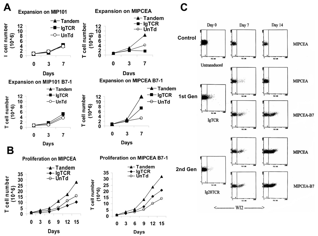
Fig. 2.
Proliferation. A. Enhanced proliferation is both CEA-dependent and Signal 2-dependent. On days specified, T cells (untransduced, IgTCR [45% modified], Tandem [15% modified]) were counted, split and mixed with an equal number of irradiated tumor cells: MIP101 (CEA neg) or MIPCEA (CEA+), without or with B7 co-expression. B. Longer term assay. Experiment as in A., except for two weeks duration and only employing CEA+ tumor targets with T cells. A and B are single experiments, repeated on three occasions with comparable results. C. Selective expansion of Tandem T cells. 1st or 2nd generation designer T cells were prepared and adjusted to very low (~2%) starting fractions. (See Methods.) T cells were co-cultured with specified tumor cells, and the fractions of modified T cells were assayed by WI2+ staining on FACS. D. Summary of results from C. Proliferative result (“Prolif”) is relative to basal expansion of activated but un-restimulated T cells.

