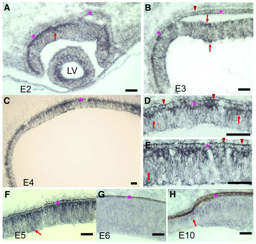
Fig. 2.
The expression of ngn3 in the developing chick retina detected with in situ hybridization. (A, B) Cross-sections of an E2 eye (A) and an E3 eye (B). Arrows points to ngn3-expressing cells in the retina. Arrowhead points to a ngn3-expressing cell in the RPE. (C) E4 retinal neuroepithelium. (D) Higher magnification of a peripheral region of E4. Arrows point to ngn3-expressing cells in the retinal neuroepithelium. Arrowheads point to ngn3-expressing cells in the RPE. (E) Higher magnification of central region of E4. Arrow points to a ngn3-expressing cell in prospective ganglion cell layer. Arrowheads point to ngn3-expressing cells in the RPE. (F) E5 retinal neuroepithelium. Arrow points to a ngn3-expressing cell in the prospective ganglion cell layer. (G) E6 central retina. (H) the peripheral retina at E10. Arrow points to a ngn3-expressing cell at the tip of peripheral retina. The RPE layer is indicated by an asterisk. LV: the lens vesicle. Scale bars: 50 µm.

