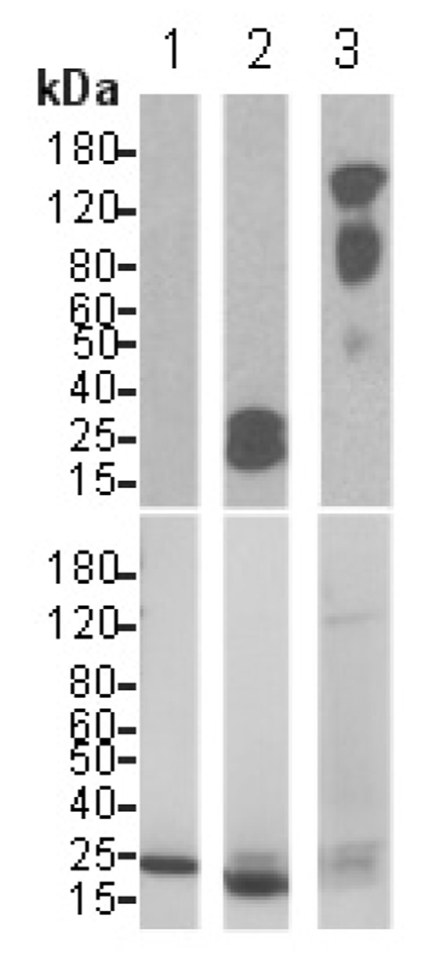
Fig. 6.
VPO1 covalently binds to heme. The experimental procedures are described as in Materials and Methods. The blot in the upper panel was developed with Pierce’s chemiluminescent substrate and chemiluminescent signal was detected by exposure to X-ray film. After extensive washing, the same blot was stained with Coomassie’s Blue and destained using standard procedures (lower panel). 1. myoglobin; 2. cytochrome c; 3. VPO1. Molecular size scale is shown on the left.

