Fig. 5.
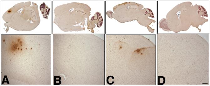
Amyloid-β deposition profile in aged BACE1xAPP animals. Brains from 13-month (A, B) and 18-month (C, D) old animals were fixed in 10% formalin, embedded in paraffin, and sectioned on a sagittal plane (10 μm thick). Scattered sections were analyzed by staining using standard protocols [17] with mAb 6E10, which detects amino acids 1-17 in the Aβ region. Shown are whole brain scans (upper) and frontal cortex brain sections (lower) of animals transgenic for both BACE1 and homozygous R1.40 YAC APP at 13 months (A) and 18 months (C). Shown are whole brain scans (upper) and frontal cortex brain sections (lower) of animals transgenic for homozygous R1.40 YAC APP alone at 13 months (B) and 18 months (D). Scale bar in D = 100 μm.

