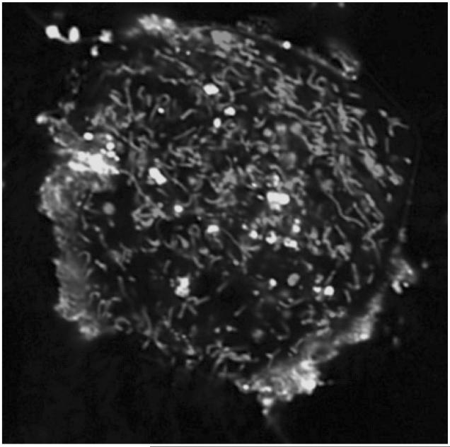
Figure 1.
Confocal fluorescence microphotograph of mouse embryonic cells co-incubated with fluorescently labeled CL (NBD-CL). Mitochondria were stained with MitoTracker® Red CMXRos. NBD-CL was delivered into cell by co-incubation with small unilamellar liposomes containing NBD-CL and 1,2-dioleoyl-sn-glycero-3-ethylphosphocholine (2:1) for 15 min at 37°C in PBS. Note that NBD-CL is not integrated into mitochondria but stays isolated in on the cell surface or sequestered in separate vacuoles. (Please see Supporting Information for an enlarged version of this figure in color.)

