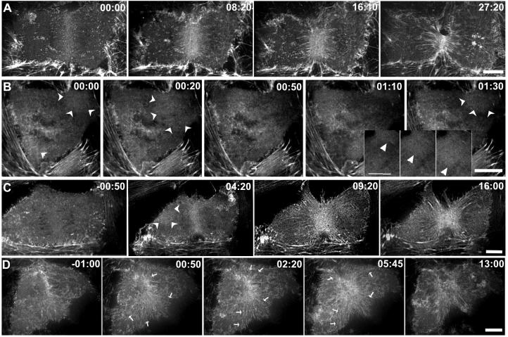
Figure 2. Microtubule disassembly induces changes in the organization of cortical actin.
LLC-Pk1 cells stably expressing GFP-actin are shown before and at various times after addition of 33μM nocodozole. Contractile ring formation and cytokinesis in a control cell (A) and cells to which nocodazole was added either within 2 minutes (B) or > 2 minutes after (C, D) anaphase onset. In (B) both contractile ring assembly and cytokinesis fail, whereas in (C, D) furrow formation and ingression proceed. Wave-like behavior of cortical actin is shown in the inset panels in (B). Arrowheads mark wave-like behavior in (C,D). In (D) actin from distal regions of the cortex flows towards the equatorial region and contributes to the contractile ring; arrows mark the direction and region contributing to flow. Time in minutes:seconds relative to the addition of nocodozole in (B,C,D). Bars = 10 μm.

