
Figure 3. Localization of GFP-RhoA in control and in nocodazole treated cells.
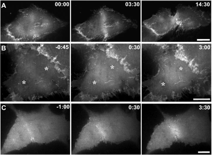
Fluorescence images of living LLC-Pk1 cells transfected with GFP-tagged C.elegans RhoA (GFP-CeRhoA). In control cells (A) and nocodazole treated cells that assemble a contractile ring (C), GFP-CeRhoA localizes to the equatorial cortex during cytokinesis. In nocodazole treated cells that fail to assemble a contractile ring (B), GFP-CeRhoA fails to accumulate at the equatorial region; asterisks mark the location of chromosomes. In both control and nocodazole treated cells GFP-CeRhoA also localizes to membranous folds between neighboring cells. Time is in min:sec. Bar = 10 μm.