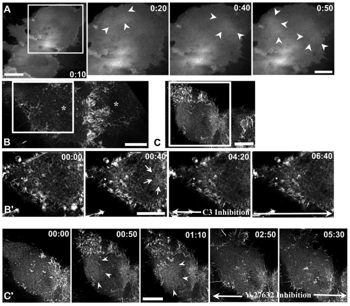
Figure 6. Rho is mislocalized and its activity is required for wave-like behavior of cortical actin in nocodazole treated cells.
(A) GFP-CeRhoA is mislocalized to the polar region in nocodazole treated cells; arrowheads mark an area of GFP-CeRhoA fluorescence in the polar cortex. (B, C) LLC-Pk1 cells expressing GFP-actin were treated with nocodazole in anaphase, and then injected with C3 (B) or treated with Y-27632 (C). White boxes indicate the region that is shown in (B’, C’). Addition of either C3 or Y-27632 suppresses wave-like behavior (B’, C’); arrows show sites where cortical actin accumulated during wave-like behavior following treatment with nocodazole. Time of addition of C3 or Y27632 is indicated by black arrow. Time is in min:sec. Bars = 10 μm.

