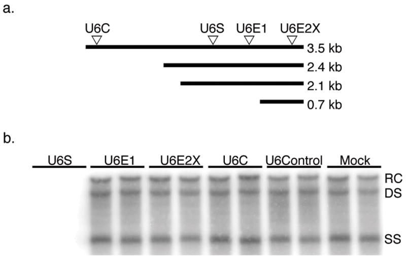
FIG. 1. Inhibition of HBV RI DNA by transfection with shRNA expressing plasmids.

(a) Arrows represent location of RNAi target sites within the HBV transcripts. (b) HBV RI DNA was extracted from HepG2 cells four days post-cotransfection with a HBV expressing plasmid and either shRNA expressing plasmid pBBU6S, pBBU6E1, pBBU6E2X, pBBU6C or negative control pBBU6Control. Cells were also transfected with HBV expression plasmid alone (Mock). HBV RI DNA was visualized by Southern blotting. Bands indicate the relaxed circular (RC), double stranded (DS), and single stranded (SS) HBV RI DNA species.
