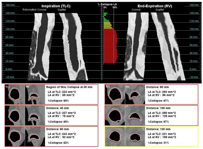
Figure 1.
Example of an analysis report; reformatted coronal and sagittal views along the trachea are shown at both inspiration (top-left) and end-expiration (top-right) breathhold; the color-coded degree of collapse is shown at each measurement point along the trachea (top-middle); examples cross-sectional views of the trachea at both inspiration levels are shown side-by-side at selected locations (bottom).

