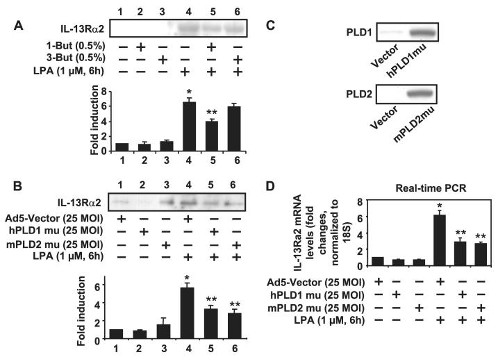
FIGURE 3. LPA-induced IL-13Rα2 gene expression and protein release was dependent on PLD in HBEpCs.
A, HBEpCs grown to ∼70–80% confluence were treated with 1-butanol (0.5%) and 3-butanol (0.5%) for 15 min. Cells were challenged with LPA (1 μM) for an additional 6 h, and then media were collected, subjected to 10% SDS-PAGE, and analyzed to a specific antibody to IL-13Rα2. This is a quantitative analysis from three independent experiments (mean ± S.D.). *, p < 0.05 versus control cells; **, p < 0.05 versus LPA treatment. B, HBEpCs grown to ∼50–60% confluence were infected with adenoviral vector containing cDNA of empty Ad5-vector, hPLD1 mutant, and mPLD2 mutant for 24 h. Cells were challenged with LPA (1 μM) for an additional 6 h, and then media were collected, subjected to 10% SDS-PAGE, and analyzed to a specific antibody to IL-13Rα2. This is a quantitative analysis from three independent experiments (mean ± S.D.). *, p < 0.05 versus adenoviral empty vector Infected cells; **, p < 0.05 versus LPA treatment. The expression of hPLD1 and mPLD2 were determined by immunoblotting with specific antibodies to PLD1 and PLD2 C and D, cells were treated as described in B. Total RNA was extracted, and IL-13Rα2 mRNA levels were determined by real-time RT-PCR with IL-13Rα2 primers. Data are the mean ± S.D. of three values. *,p < 0.05 versus adenoviral empty vector-infected cells; **, p < 0.05 versus LPA treatment. MOI, multiplicity of infection.

