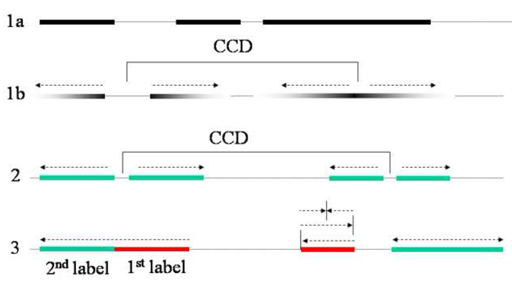
Figure 1. Analysis of initiation and elongation of replication using pulse labeling of DNA with subsequent stretching.
1A) 3H thymidine labeling allows measuring lengths of labeled tracks in stretched DNA after exposure to photo emulsion. 1B) A high specific activity pulse of 3H thymidine followed by a low specific activity chase provides information about direction of replication fork movement. Replicon center to center distance (CCD) can be measured as a distance between centers of symmetry of pairs of divergent forks. 2) Synchronization of cells early in S phase by aphidicolin and pulsing with BrdU 5 minutes after release from aphidicolin visualizes replication bubbles in stretched DNA as pairs of tracks with small gaps of unlabeled DNA in between. CCD would correspond to a distance between these small gaps. 3) Double labeling with consecutive pulses of IdU and CldU allows distinguishing ongoing forks, terminated forks and newly fired forks, and shows direction of movement of ongoing forks.

