
Figure 4.
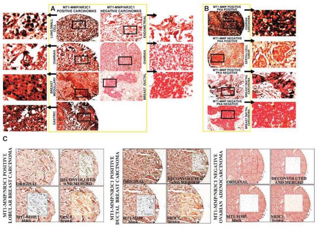
Representative immunostaining of MT1-MMP, NR3C1, and PKAα in human cancer specimens arranged in TMAs. A, MT1-MMP and NR3C1 double staining. B, MT1-MMP and PKAα double staining. TMAs were stained with the antibodies to PKAα and NR3C1 (diaminobenzidine, brown) and with the antibody Ab815 to MT1-MMP (gray black) and counterstained with Nuclear red. The areas marked with squares were enlarged to facilitate the visual analysis of the colors. Bar, either 50 or 100 μm. C, analysis of the images. The selected regions (boxed) were subjected to the image analysis system using Scanscope-HT (Aperio Technology). The black and brown colors of the original image were separated using a color deconvolution algorithm (“deconvoluted and merged” panels). The separated colors are shown in the “MT1-MMP, black” and the “NR3C1, brown” panels. There is an obvious colocalization of MT1-MMP and NR3C1 in the breast carcinoma biopsies, whereas the ovarian carcinoma sample was negative in both MT1-MMP and NR3C1 markers. Original magnification, 300×.