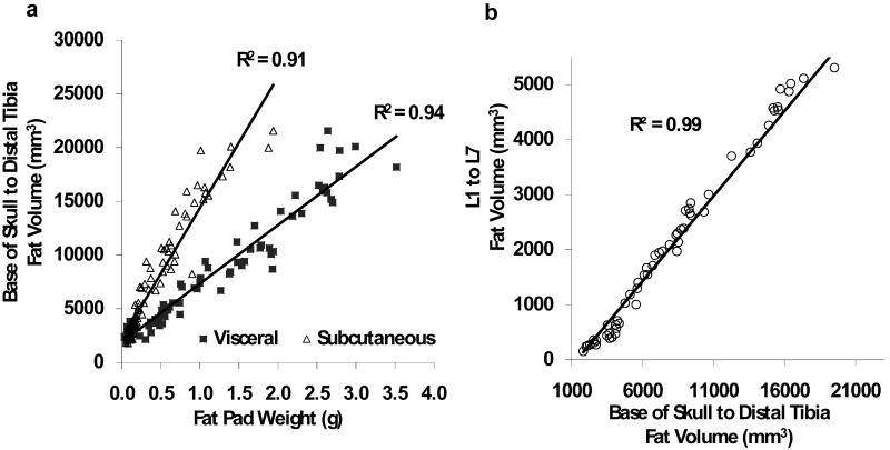
Figure 4.
(a) Total fat volume (from the base of the skull to the distal tibia) determined by in vivo microCT was highly correlated with both the visceral and subcutaneous tissue weight of the fat pads harvested at sacrifice (n=90). (b). A scan of the abdominal region reduced the scan time by two-thirds. Despite the much smaller region, fat volume of the abdomen (spanning between L1 and L5 vertebrae) was highly correlated with total fat volume of the entire mouse body (n=45).

