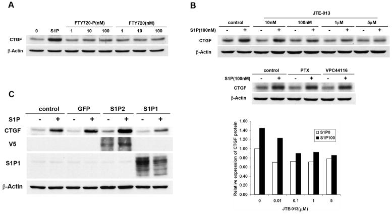
FIGURE 2.
S1P-induced CTGF expression was mediated by S1P2. A. WiT49 cells were serum starved for 24 h and then treated with 100 nM S1P or different concentrations of FTY720-P or FTY720 for 1 h before western blot analysis was done. B. WiT49 cells were pretreated with S1P2 antagonist JTE-013 (10 nM, 100 nM, 1μM, 5 μM), S1P1 antagonist VPC44116 (1 μM) or Gi protein inhibitor PTX (400 ng/ml) for 0.5 h after serum starvation and then stimulated with 100 nM S1P for another 1 h before western blot analysis was done. The relative expression of CTGF protein in cells treated with or without JTE-013 was normalized to that in control cells without JTE-013 or S1P treatment which was regarded as 1. S1P0 and S1P100 mean no S1P and 100 nM S1P treatment, respectively. C. WiT49 cells were infected with adenovirus overexpressing S1P2, S1P1 or GFP with MOI 100. After 16–24 h, cells were serum starved for 24 h and then stimulated with 100 nM S1P for another 2 h before western blot analysis was done.

