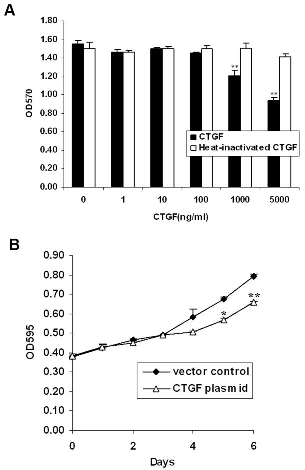
FIGURE 5.
CTGF inhibited cell proliferation in WiT49 cells. A. WiT49 cells were plated to 96-wells. After attachment, they were serum starved for 24 h and then incubated with different concentrations of human recombinant CTGF or heat-inactivated CTGF by being boiled for 10 min for an additional 48 h before the MTT assay was conducted. Data are the mean±SD of triplicates. **, P < 0.01 versus without CTGF. B. WiT49 cells were transfected with either CTGF plasmid or vector control and cultured for indicated days followed by MTT assay. Growth rates were compared between the CTGF plasmid and vector control transfected cells. *, P < 0.05, **, P < 0.01 versus vector control.

