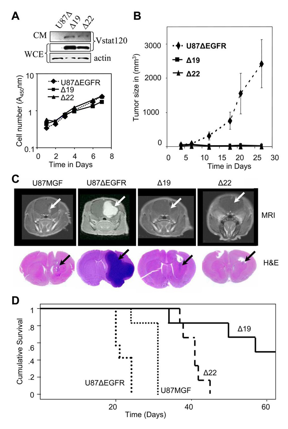
Figure 2. Vstat120 expression suppresses subcutaneous and intracranial tumor growth of U87ΔEGFR cells despite the pro-angiogenic stimulus provided by EGFRvIII.
A. Characterization of U87ΔEGFR and Vstat 120 expressing clones Δ19 and Δ22. Upper Panel shows western blot analysis of whole cell extract (WCE) and conditioned medium (CM) from U87ΔEGFR cells (U87Δ) and derived clones Δ19 and Δ22, which stably express Vstat120. Lower panel shows in vitro proliferation rates of U87ΔEGFR cells, and Vstat120 expressing clones Δ19 and Δ22, as measured using the crystal violet assay. Expression of Vstat120 does not alter the in vitro proliferation rates of these cells.
B. Subcutaneous growth of U87ΔEGFR and Vstat120-expressing clones in nu/nu mice. U87ΔEGFR and derived clones stably expressing Vstat120 (Δ19 and Δ22) were injected subcutaneously into mice (n=6) and the tumor volume for the indicated clones was plotted as a function of time. Note the strongly decreased tumor growth of clones expressing Vstat120.
C. Relative growth of U87MGF, U87ΔEGFR and Vstat120-expressing clones in rat brains. Upper panels show representative images of the MRI scans of individual rat brains 14 days after intracranial implantation of 106 tumor cells. The presence of glioma is detected through the bright areas (white arrows) of contrast enhancement from the MRI contrast agent (Gd-DTPA). Note the small tumor in U87MGF cells, large tumor in U87ΔEGFR cells and barely detectable minimal tumors in clones Δ22 and Δ19. The lower panel shows corresponding histopathological brain sections stained with H&E. Tumor growth is visible as a darkly stained area (black arrow).
D. Survival curves of rats implanted with U87MGF, U87ΔEGFR and Vstat120 expressing clones, Δ 19 and Δ22. 1 × 10⁶ cells were implanted stereotactically in the brain of athymic nu/nu rats. Rats implanted with U87ΔEGFR cells had the shortest survival time due to the very angiogenic and aggressive nature of these tumors. Vstat120 expressing clones Δ19 and Δ22, showed a significant improvement in their survival compared to the U87ΔEGFR and control parental U87MGF cells (p<0.05).

