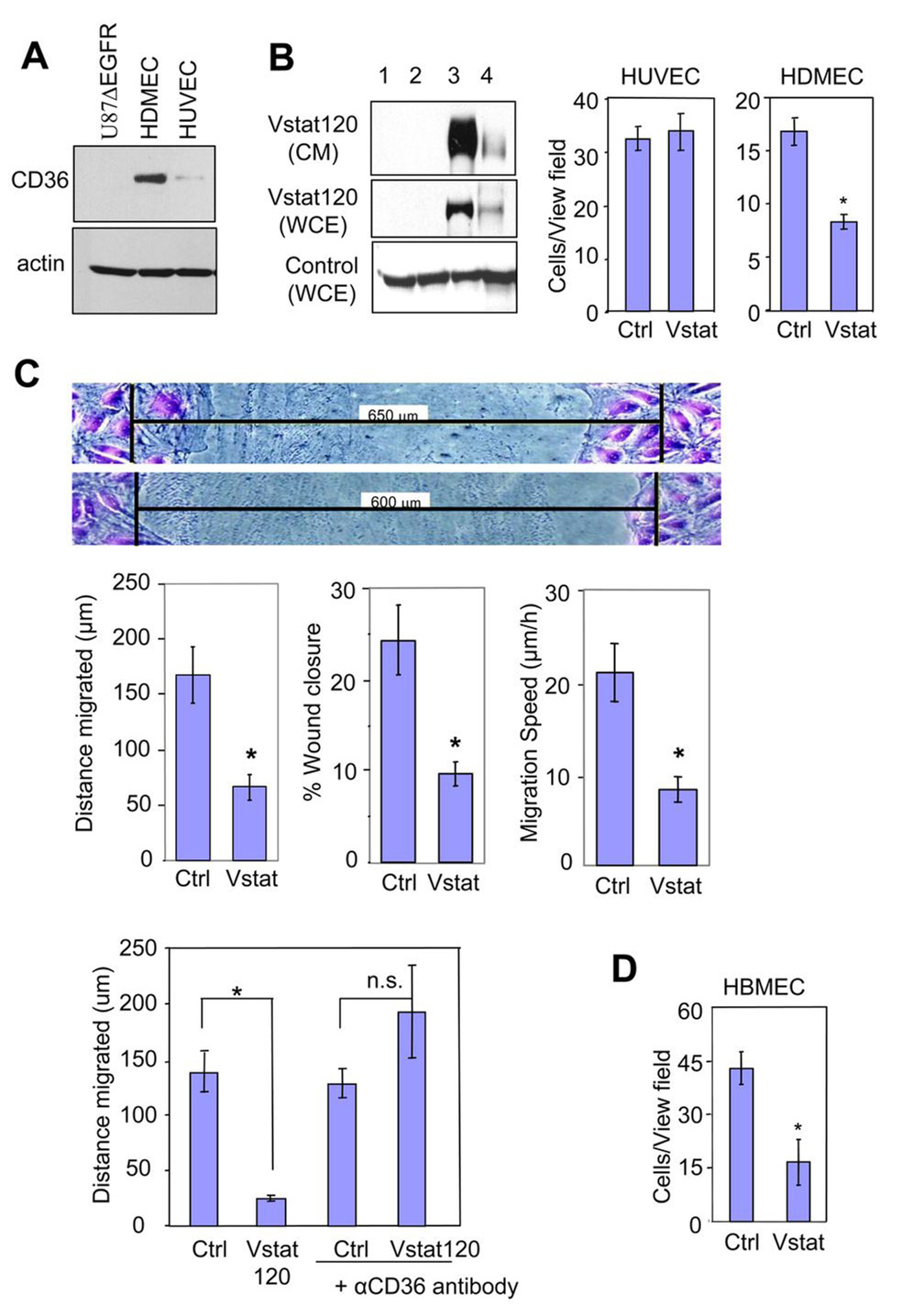
Figure 4. Vstat120 inhibits endothelial cell migration in a CD36-dependent fashion.
A. Western blot analysis for expression of CD36 in U87ΔEGFR, HDMEC, and HUVEC cells, respectively.
B: Production of secreted Vstat120 by transient transfection in 293 cells (Left). The cells (80% confluent) were left untreated (lane 1) or were transfected with either control pcDNA3.1lacZ vector (lane 2) or Vstat120 expression vector pcDNA3.1Vstat120-myc/his (lane 3). Vstat120 produced by cells transfected with full length BAI1 expression vector was utilized as a size control (lane 4). CM was collected in serum-free media for 48hrs and used in experiments B–D below. WCE, whole cell extract.
Transwell migration assays (Right) examining the migration of HUVEC and HDMEC in the presence of CM from control or Vstat120 containing CM from 293 cells. The number of cells that migrated to the bottom of the chamber after 8 hrs was quantified as described in materials and methods. Note that Vstat120 containing CM reduces the migration of HDMEC, but not HUVEC.
C. Scratch-wound migration assays. Confluent HDMECs were wounded, treated with CM prepared as in B. and the endothelial cells allowed to migrate for 8 hrs, then fixed and stained with crystal violet. Shown are representative pictures of migrated cells (Top panel). The black bars indicate initial wound width in micrometers. Distance of migration, percentage of wound closure, and speed of migration was quantified (Middle panel). The experiment was repeated twice with similar results. Data are expressed as mean +/− SEM; n=6 for each condition; * p<0.01 compared to Vstat120.
CD36 function-blocking antibody prevents Vstat120 anti-angiogenic function (Bottom panel). HDMECs were wounded, then either left untreated or treated with anti-CD36 function-blocking antibody at 10 µg/mL for 30 min. The cells were next treated with CM (as above) for 30 min, followed by treatment with 10% serum to induce cell migration. Final wound width was measured after 8 h and the distance migrated was calculated.
D. Transwell assay examining the migration of HBMEC in the presence of CM from U87MGD (Ctrl) and Vstat 120 expressing U14 cells (Vstat).
Data is mean ± SEM. n=3 for each condition. * p<0.05 and n.s. not significant by Student’s T test.

