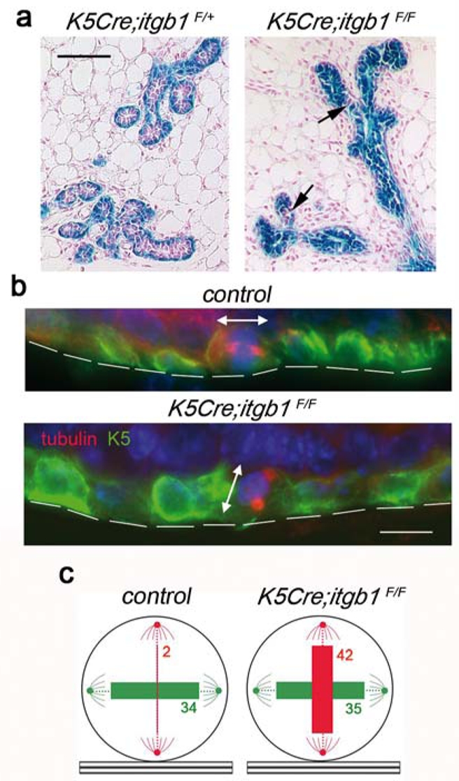
Figure 4. Altered orientation of the basal cell division axis in K5Cre;itgb1F/F mammary epithelium.

(a) Whole-mount X-gal staining of the K5Cre;itgb1F/+ and K5Cre;itgb1F/F outgrowths developed in 7.5-dpc host. Arrows show LacZ-negative (pink) cells. (b) Dividing basal cells in the ducts formed by control and K5Cre;itgb1F/F epithelium. Double immunofluorescence staining with anti-K5 and anti-β-tubulin antibodies. Basement membrane position is marked by discontinuous lines, double-headed arrows indicate division plane. (c) Position of ductal basal cell division plane in the mammary outgrowths developed in host mice at 7.5 dpc. The numbers in green and red correspond to the numbers of cells dividing parallel and perpendicular to the basement membrane, respectively. The thickness of colored bars is proportional to the cell number. Cell counts obtained for each of the four mice used for the analysis are presented in Supplementary Information, Table 1. Scale bars, 100 μm (a), and 75 μm (b).
