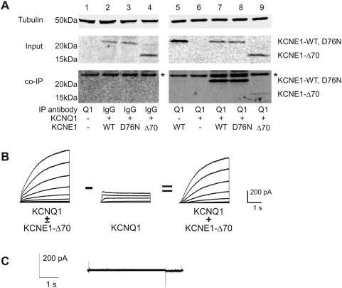
Figure 5.
A) Co-immunoprecipitation of wild-type and mutant KCNE1 with KCNQ1. KCNQ1 (Q1) was expressed with KCNE1 (E1), E1-D76N, or E1-Δ70. KCNQ1 was immunoprecipitated with goat anti-KCNQ1 antibody (Santa Cruz), and western blot analysis performed with mouse anti-FLAG antibody (Sigma). The asterisks (*) indicate a row of nonspecific bands that appeared in every lane, including control lanes. For controls, lane 1 shows results from untransfected cells, lanes 2–4 from cells transfected with KCNQ1 and KCNE1 (WT, D76N, or Δ70 as indicated) and pulled down with control IgG. B) Digital subtraction of Q1 current from mixed Q1-E1 current (E1-Δ70 mutant). Left tracing shows the currents obtained from a cell transfected with a 1∶1 ratio of KCNQ1∶KCNE1-Δ70 plasmids. Middle tracing shows a pure KCNQ1 current scaled to match the amplitude of the initial rapid current deflection seen in the mixed current to the left. Right side tracing shows the resulting digital subtraction of the two currents resulting in a current with slow sigmoidal activation characteristic of IKs. C) Voltage clamp tracing from cells transfected with KCNE1-Δ70 plasmid alone demonstrating that the mutant KCNE1 did not induce any other conductances on its own.

