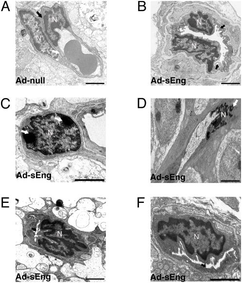
Figure 7. Ultrastructure of retinal vasculature following inhibition of TGF-β.
(A) TEM micrograph of a microvessel in the ganglion cell layer from a retina of a control mouse expressing Ad-null (day 14). Nuclei of an EC and pericyte are apparent, with a defined basement membrane (arrow). (B)–(F) TEM micrographs of the retinal vessels from mice expressing Ad-sEng. (B) The lining of some ECs appeared ‘ruffled’ with finger-like processes protruding into the luminal space and multiple vacuoles within the cytoplasmic space (arrow). Nuclear condensation characteristic of apoptosis was apparent in some (C) pericytes and (D) ECs (arrows). (E) (F) Numerous vessels in the inner retinal layers displayed significant reductions in luminal diameter (arrows). (A)–(F) Scale = 2 µm.

