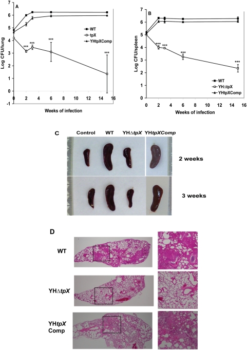
Figure 3. Growth and survival of YHΔtpx in the lungs and spleen of BALBc mice.
Mice were infected with 3 × 10⁵ bacteria. At different time points the infected mice were sacrificed and the numbers of bacteria in the lung (A) or spleen (B) were measured. The results for each time point are the means and SDs of four mice in each experimental group. The experiments have been reproductively repeated twice with similar CFU counts in lungs and spleens. Statistical significance was determined by Student's t test (***, P<0.0001). C. Changes in spleen gross anatomy after infection with the WT, the mutant and the complemented strains. Spleens were collected at 2 and 3 weeks after infection. D. Lung histology of mice infected with the WT, the mutant and the complemented strains. Histopathological examination was performed using three mice in each group. Three sections from each mouse were examined. The images shown are representative of lung sections from three animals in each experimental group. Enlarged images of the boxed regions on the left panel (magnification, 4×) are shown on the right panel (magnification, 10×).

