
FIG. 1.
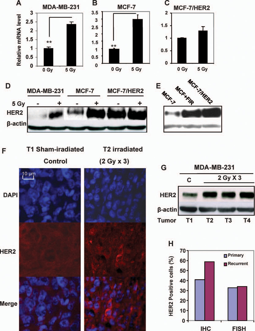
HER2 expression was induced in breast cancer cells by γ radiation. Panels A–C: HER2 mRNA levels were increased in MDA-MB-231 (panel A) and MCF-7 (panel B) breast cancer cells but not in MCF-7/HER2 cells (panel C). Total RNA purified from cells 24 h after exposure to 5 Gy of γ rays (n = 3; mean ± SE; **P < 0.01). Panels D and E: HER2 protein levels were enhanced in irradiated (5 Gy γ rays) MDA-MB-231 and MCF-7 cells (panel D) as well as in the radioresistant population that survived long-term fractionated irradiation (MCF+FIR; panel E) (21) but not in HER2-overexpressing MCF-7/HER2 cells measured by Western blot analysis. Panel F: HER2 expression was induced in irradiated MDA-MB-231 xenograft tumors (3 × 2 Gy; total tumor dose 6 Gy) detected by HER2 immunohistochemistry (red) with DAPI nuclear staining (blue) 24 h after irradiation (T1 = sham-irradiated control; additional HER2 immunohistochemistry data can be found in Supplementary Fig. S1). Panel G: Western blot of HER2 in MDA-MB-231 control (C) and irradiated xenografts 24 h after irradiation. Panel H: Increased frequency of HER2-positive breast cancer cells in human recurrent invasive breast cancers compared to primary tumors analyzed by immunohistochemistry (IHC) and FISH.