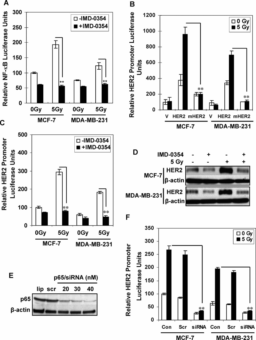
FIG. 3.
NF-κB is required for radiation-induced HER2 expression. Panel A: MCF-7 and MDA-MB-231 cells transfected with NF-κB-driven luciferase reporters were treated with IMD-0354 (2 µM) or DMSO for 5 h before exposure to 5 Gy or sham irradiation. Luciferase activities were determined 24 h after irradiation (n = 3; mean ± SE; **P < 0.01). Results are shown as the percentage of the value for untreated MCF-7 cells. Panel B: pGL2 luciferase plasmid (V), HER2-controlled luciferase reporter (HER2), or HER2-controlled luciferase reporter with deleted NF-κB binding site (mHER2) was transfected into MCF-7 and MDA-MB-231 cells. Luciferase activity was measured 24 h after exposure to 5 Gy or sham irradiation (n = 3; mean ± SE; **P < 0.01). Panels C and D: MCF-7 and MDA-MB-231 cells transfected with HER2 luciferase reporters were treated with IMD-0354 (2 µM) or DMSO for 5 h before exposure to 5 Gy or sham irradiation. Luciferase activities were determined 24 h after irradiation (panel C; n = 3; mean ± SE; **P < 0.01). Panel D: Western blot analysis of HER2 levels 24 h after irradiation. Panels E and F: Western blot analysis of MCF-7 cells treated with scramble siRNA or p65 siRNA for 60 h (panel E; lip = transfectants reagent only; scr = 20 nM scrambled siRNA; siRNA = p65 siRNA), and luciferase activity was measured 24 h after irradiation. Results are shown as a percentage of the value for untreated MCF-7 cells (panel F; n = 3; mean ± SE; **P < 0.01).

