
FIG. 6.

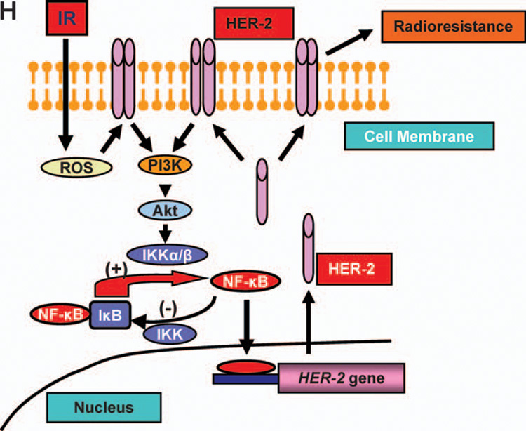
Radiosensitization by HER2 siRNA. Panel A: Inhibition of HER2 expression in MCF+IR and MCF-7/HER2 cells by 20 nM HER2 siRNA (lip =transfection reagent; scr =20 nM scrambled siRNA; siRNA = HER2 siRNA). Panels B and C: Clonogenic survival of MCF-7/HER2 (panel B) and MCF+IR (panel C) cells treated with HER2 siRNA (20 nM for 60 h) and then irradiated (points, mean; n = 3; bars, SE; **P < 0.01, *P < 0.05 compared to the scramble siRNA control). Panel D: Inhibition of HER2 expression in radioresistant (C4, C5) and radiosensitive (C2) MDA+FIR cells treated with HER2 siRNA or scrambled siRNA (20 nM for 60 h). Panels E–G: Clonogenic survival of radiosensitive C2 (panel E) and radioresistant C4 (panel F) and C5 (panel G) cells treated with HER2 siRNA (20 nM for 60 h before irradiation; n = 3; mean ± SE; **P < 0.01). Panel H: Schematic representation of radiation-induced loop-like HER2-NF-κB-HER2 pathway in radiation-induced adaptive resistance. IR, radiation.