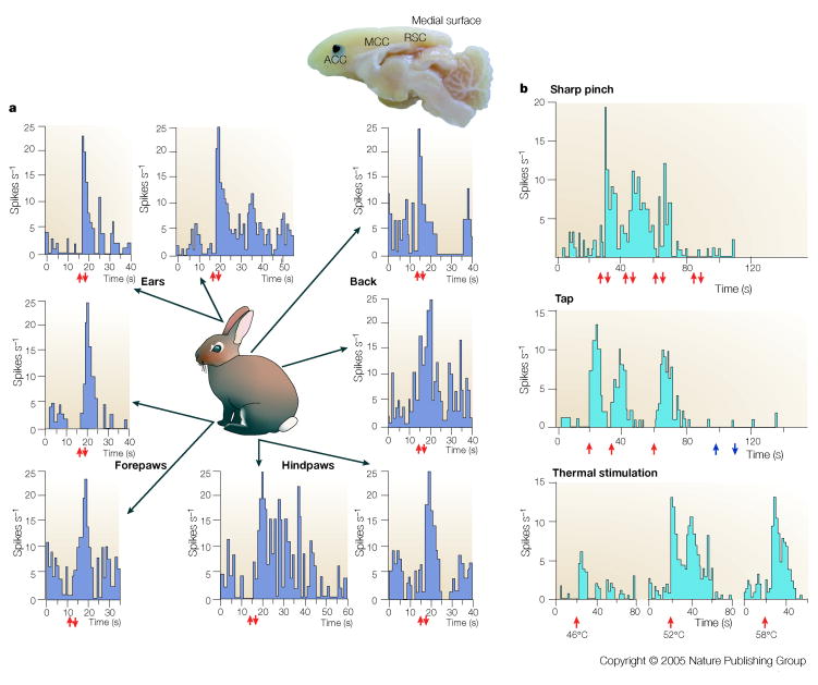
Figure 2. Nociceptive Cingulate Neurons.
Medial surface of the rabbit brain showing its cingulate regions (ACC, MCC, RSC) and the location of a high density of nociceptive neurons (black) and a low-moderate density of such neurons (grey). (The rabbit does not have PCC areas 23 and 31 like primates.) The rasters display neuron spike discharges for two neurons and the arrows show stimulus onset (↑) and offset (↓). Noxious mechanical stimulation with a serrated forceps evoked a response of the neuron on the left regardless of where the stimulus was applied to the skin and demonstrates that no one neuron in ACC can determine where on the body a noxious stimulus is located. The neuron on the right is a multimodal nociceptive neuron because it responds briskly to both noxious mechanical and heat stimulation. Although the unit did respond to tap stimulation, light brushing of the skin (open arrows) failed to evoke a response. These multimodal nociceptive units provide little information about the characteristics of particular noxious stimuli.

