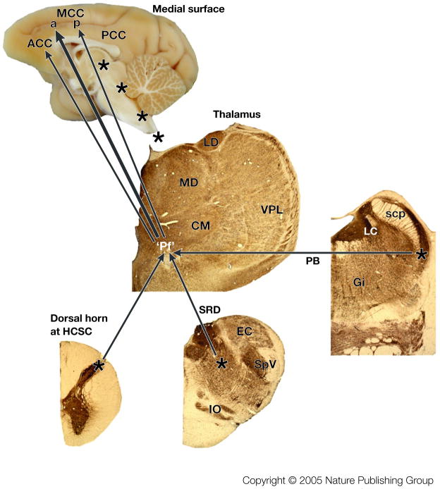
Figure 4. Nociceptive afferents to cingulate cortex.
Three sources of nociceptive inputs to “Pf” arise from lamina I of the spinal cord, subnucleus reticularis dorsalis (SRD) and the parabrachial nucleus (PB). The four asterisks along the medial surface of the monkey brain show the levels at which the thalamic, PB, SRD, and high cervical spinal cord sections were photographed. These are immunohistochemical sections for an antibody to microtubule associated protein 2 (MAP2) which label neuron dendrites and they were counterstained with thionin because some neurons do not express this antigen. The Pf nucleus is enclosed in quotation marks because it is a representative of the MITN of which there are many nuclei that receive nociceptive spinothalamic inputs and project in turn to cingulate cortex (see BOX #2). We believe the greatest density of nociceptive inputs is to aMCC as shown with the large arrow, while more modest projections are to ACC and pMCC. Abbreviations: CM, centre medianum thalamic nucleus; EC, external cuneate nucleus; Gi, gigantocellular reticular nucleus; IO, inferior olive; LC, locus coeruleus; LD, laterodorsal thalamic nucleus; MD, mediodorsal thalamic nucleus; scp, superior cerebellar peduncle; SpV, spinal nucleus and tract of the trigeminal nerve; VPL, ventral posterolateral thalamic nucleus.

