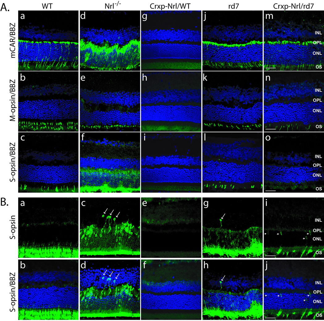
Figure 3. Expression of cone-specific markers and targeting of photoreceptors to the ONL.
(A) Superior, and (B) inferior regions of the retina showing staining for cone arrestin (mCAR), S-opsin, and M-opsin antibodies (as shown on the left). Mouse strains are indicated. Compared to WT (B: a–b) and Crxp-Nrl/WT (B: e–f), targeting of S-cones (arrows) to the ONL is perturbed in Nrl−/− (B: c–d) and rd7 retinas (B: g–h), and S-opsin positive nuclei are present in the INL. S-cone staining (arrowheads) in the Crxp-Nrl/rd7 retinas (B: i–j) is observed in cells closest to the outer plexiform layer. OS, outer segments; ONL, outer nuclear layer; OPL, outer plexiform layer; INL, inner nuclear layer; BBZ, bisbenzamide. Scale bar: 25 µm.

