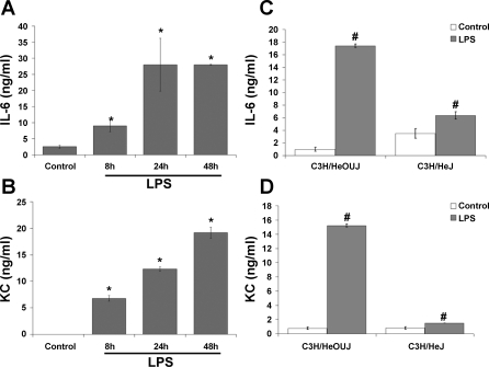
Fig. 4.
LPS induces secretion of IL-6 and keratinocyte-derived chemokines (KC) in intestinal MF. Cells were treated as in Fig. 2. Supernatants were collected and analyzed by ELISA for IL-6 (A) and KC (B). Data represent means ± SD and are representative of 4 independent experiments. *P < 0.05 vs. control. C and D: IL-6 and KC secretion, respectively, after 24 h of LPS stimulation in MF derived from LPS-resistant C3H/HeJ or wild-type syngeneic C3H/HeOUJ mice. #P < 0.05 vs. control of the same cell type.

