Fig. 4.
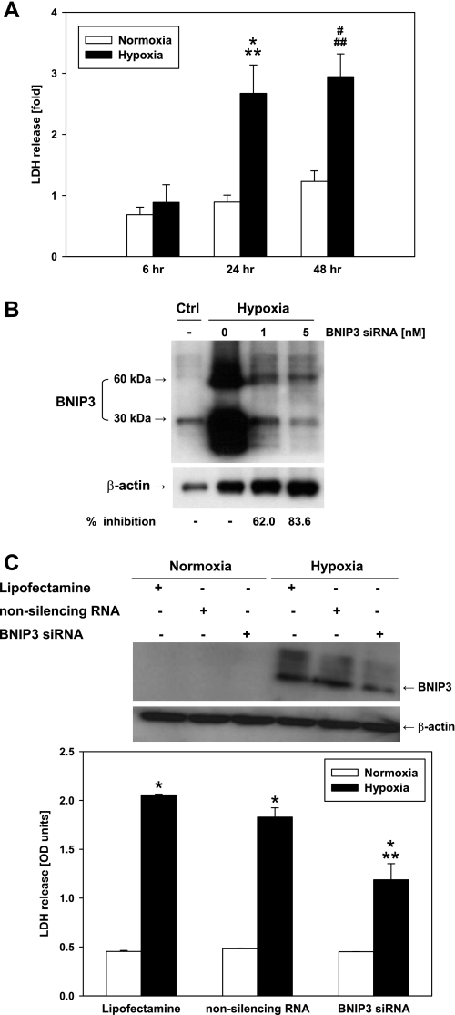
BNIP3 small interference (si)RNA protects hepatocytes from hypoxia-induced cell injury. A: freshly isolated mouse hepatocytes were exposed to 6, 24, and 48 h of hypoxia (1% O2), and viability was assessed by LDH release. Results are means ± SE (n = 9–16). *P < 0.005 vs. normoxia, 24 h. **P < 0.05 vs. hypoxia, 6 h. #P < 0.01 vs. normoxia, 48 h. ##P < 0.005 vs. hypoxia, 6 h. Data were analyzed by 1-way ANOVA followed by Tukey's test. B: after transient transfection with BNIP3 siRNA (1 and 5 nM), primary mouse hepatocytes were exposed to 24 h of hypoxia and the total protein was isolated. A representative Western blot showing the knockdown of BNIP3 gene is shown (n = 3). The inhibition of BNIP3 protein expression was calculated using the densitometric analysis for the 30-kDa band. Ctrl, control. C: freshly isolated mouse hepatocytes were treated with Lipofectamine, nonsilencing (scrambled) RNA, and BNIP3 siRNA (5 nM) for 24 h of hypoxia (1% O2), and viability was assessed by LDH release. OD, optical density. *P < 0.001 vs. normoxia. **P < 0.001 vs. hypoxia (Lipofectamine and nonsilencing RNA). Data were analyzed by 1-way ANOVA followed by Tukey's test. Representative Western blot (top) shows BNIP3 protein expression under the same experimental conditions as described above.

