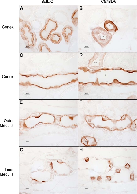
Fig. 1.
Light microscopic examination of Rh C glycoprotein (Rhcg) immunoreactivity using antibody A in Balb/c and C57BL/6 mouse kidney. A: connecting segments in Balb/c mouse kidney. Strong apical and faint basolateral immunolabel are present. B: connecting segment (*) and distal convoluted tubule (DCT; **) in a C57BL/6 mouse kidney. Both apical and basolateral Rhcg immunolabel are easily evident. C: cortical collecting duct (CCD) in a Balb/c mouse kidney. Strong apical and weak basolateral immunolabel are present. D: CCD from a C57BL/6 mouse kidney. Both strong apical and strong basolateral immunolabel are present in the CCD (*). A DCT with lesser intensity apical and basolateral Rhcg immunolabel is also demonstrated (**). E: outer medullary collecting duct (OMCD) from a Balb/c kidney. Apical immunolabel is easily evident; basolateral immunolabel is of lesser intensity. F: OMCD in a C57BL/6 mouse kidney. Both apical and basolateral Rhcg immunolabel are relatively strong. G: inner medullary collecting ducts (IMCD) in the Balb/c kidney. Strong apical and weak basolateral immunolabel are evident. H: IMCD from a C57BL/6 kidney, where both strong apical and basolateral Rhcg immunolabel are present. The same dilution of primary antibody, 1:40,000, was used in all panels. Results are representative of findings in at least 6 mice.

