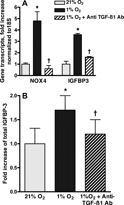
Fig. 7.
Anti-TGF-β1 antibody (Ab) inhibits hypoxic expression of NOX4 and IGFBP-3. HPASMC were incubated in 1% or 21% FiO2 for 24 h with and without anti-TGF-β1 antibody (50 ng/ml). A: real-time PCR of reverse-transcribed mRNA was performed using IGFBP-3 and NOX4 primers. Values were normalized to 18S and expressed as fold induction over normoxic control. Results are representative of 3 independent experiments. B: IGFBP-3 protein in HPASMC culture media was quantified by ELISA. Results are expressed as fold increase compared with normoxic cells and represent the means of at least 3 samples ± SD. *P < 0.05 1% O2 vs. 21% O2; †P < 0.05, 1% O2 vs. 1% O2 + anti-TGF-β1 antibody.

