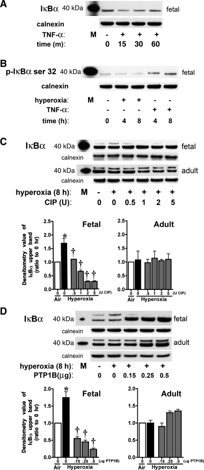
Fig. 3.
IκBα phosphorylation occurs in fetal cells exposed to hyperoxia. Fetal and adult lung fibroblasts were exposed to TNF-α or hyperoxia. A: representative Western blot showing levels of cytosolic total IκBα in control and TNF-α-treated (20 ng/ml for 15, 30, or 60 min) fetal cells, with 40-kDa marker and calnexin as loading control. M, marker. B: representative Western blot showing cytosolic levels of IκBα phosphorylated on serine 32 (p-IκBα) in control, hyperoxia-exposed (4 or 8 h), and TNF-α-treated (20 ng/ml for 4 or 8 h) fetal cells, with 40-kDa marker and calnexin as loading control. C: representative Western blots showing IκBα pattern after incubation of cytosolic fractions from hyperoxia-exposed fetal and adult cells with 0.5–5 U of calf intestinal phosphatase (CIP), with 40-kDa marker and calnexin as loading control (top), and densitometric evaluation of fold change in the upper band of IκBα after exposure to CIP (bottom). Values are means ± SE of 3 independent experiments for each group. *P < 0.001 vs. time 0. †P < 0.001 vs. 8 h without CIP. D: representative Western blots showing IκBα pattern after incubation of cytosolic fractions from hyperoxia-exposed fetal and adult cells with 0.15–0.5 μg of a tyrosine-specific phosphatase (PTP1B), with 40-kDa marker and calnexin as loading control (top), and densitometric evaluation of fold change in the upper band of IκBα after exposure to PTP1B (bottom). Values are means ± SE of 3 independent experiments for each group. *P < 0.001 vs. time 0. †P < 0.001 vs. 8 h without PTP1B.

