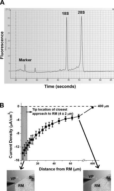
Fig. 2.
Quality of isolated RNA and relationship between position of vibrating probe and current density from Reissner's membrane. A: representative electropherogram showing high quality of total RNA obtained from Reissner's membrane. Sharp peaks representing 18S and 28S rRNA demonstrate high quality of RNA. B: distance-response curve of current density from Reissner's membrane detected by vibrating probe. Probe was nominally placed 4 μm from apical surface of the epithelium, with an estimated precision of ±2 μm (hatched vertical rectangle). Measurements therefore had variation that was intrinsic not only to different tissue samples, but also to precision of electrode placement. Current density was ∼0 at 400 μm from the tissue, the distance taken as reference. Values are means ± SE (n = 12).

