Fig. 6.
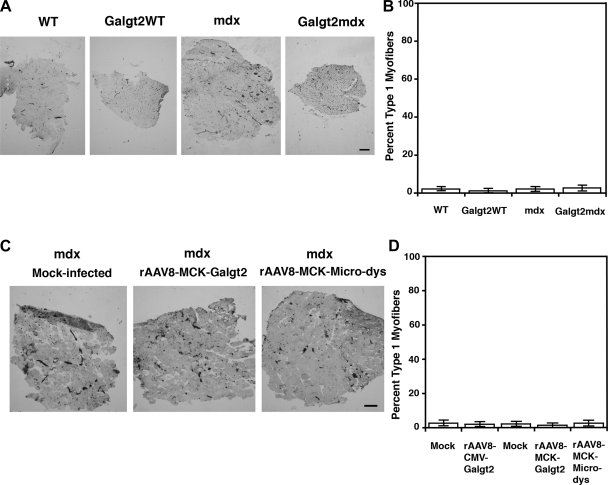
Fiber type composition of EDL muscles in all experimental conditions. A: fiber type composition of the EDL muscle of WT, Galgt2WT, mdx, or Galgt2mdx 2-mo-old muscles was determined using ATPase immunohistochemical staining at pH 4.2 to define slow (type 1) fibers, which are black, and fast (type II) subtypes of myofibers, which are not stained. Blood vessels and capillaries are also stained black with this staining method. B: quantitation of fiber type composition showed no change between the four groups. C: fiber type composition of mdx muscles treated with rAAV8-CMV-Galgt2(mouse), rAAV8-MCK-Galgt2(human), rAAV8-MCK-microdystrophin(human), or appropriate mock-infected controls. D: quantitation of fiber type composition showed no change between the five groups. Bar is 200 μm. Errors are SE for n = 6 muscles per condition.

