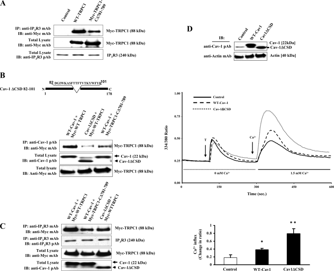
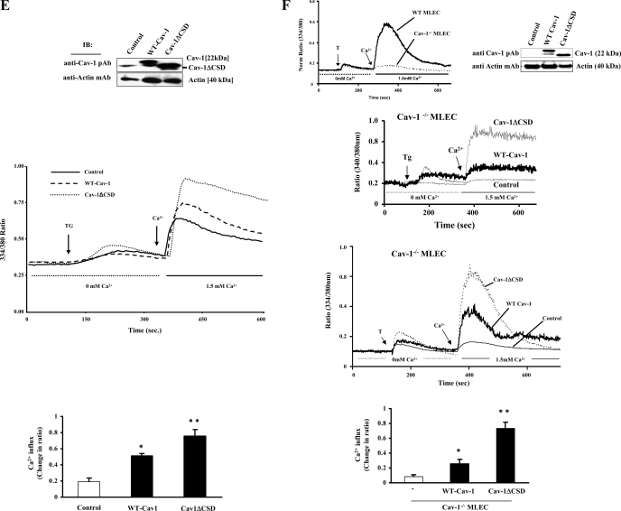
Fig. 3.
A: caveolin-1 (Cav-1) scaffold domain (CSD)-binding motif deletion of TRPC1 COOH-terminal impairs TRPC1 binding with inositol 1,4,5-trisphosphate receptor 3 (IP3R3). HEK-293 cells were transfected with Myc-WT-TRPC1 or Myc-TRPC1-CΔ781-789 expression constructs (see details in materials and methods). At 48 h after transfection, cells were harvested and the proteins extracted were subjected to immunoprecipitation using anti-IP3R3 mAb. Immunoprecipitate was resolved in a SDS-PAGE and immunoblotted with anti-Myc mAb to determine TRPC1 binding (top). Total cell lysates were immunoblotted with anti-Myc mAb (middle) or anti-IP3R3 pAb (bottom). Results are representative of 3 independent experiments. B: Cav-1ΔCSD expression reduces TRPC1 binding to Cav-1. HEK-293 cells were cotransfected with different combinations of TRPC1 and Cav-1 expression constructs. At 48 h after transfection, cells were lysed and immunoprecipitated with anti-Cav-1 pAb and blotted with anti-Myc mAb (top). Total cell lysates were immunoblotted with anti-Cav-1 pAb (middle) and anti-Myc-mAb (bottom). The experiment was repeated 4 times, and results from representative experiments are shown. C: Cav-1ΔCSD expression reduces TRPC1 binding to IP3R3. HEK-293 cells were cotransfected with different combinations of TRPC1 and Cav-1 expression constructs. At 48 h after transfection, cells were lysed and immunoprecipitated with anti-IP3R3 mAb and blotted with anti-Myc mAb (top). Total cell lysates were immunoblotted with anti-IP3R3 pAb (second from top), anti-Myc-mAb (third from top), or anti-Cav-1 pAb (bottom). The experiment was repeated 3 times, and results from representative experiments are shown. D and E: Cav-1ΔCSD mutant expression augments agonist-induced Ca2+ store release-induced Ca2+ influx in HMEC and HEK-293 cells. D: HMEC were transfected with the WT-Cav-1 or Cav-1ΔCSD expression constructs. At 48 h after transfection, cells were lysed and immunoblotted with anti-Cav-1-pAb to determine Cav-1 protein expression. The membrane was then stripped and reprobed using anti-β-actin mAb (bottom). Next, HMEC transfected with either WT-Cav-1 or Cav-1ΔCSD expression constructs were used for thrombin-induced intracellular Ca2+ influx measurements (see details in materials and methods). At 48 h after transfection, cells were loaded with Fura-2 AM and placed in Ca2+ and Mg2+-free HBSS and stimulated with thrombin (50 nM) to measure Ca2+ store depletion. After return of [Ca2+]i to baseline levels, 1.5 mM CaCl2 was applied to the extracellular medium to induce Ca2+ influx. Arrow indicates the time at which cells were stimulated with thrombin or Ca2+ was added. Experiments were repeated at least 4 times. A representative profile is shown. Change in peak fluorescence ratio (334/380) for the Ca2+ influx in control and WT-Cav-1- or Cav-1ΔCSD-expressing cells was compared. *P < 0.05, significantly different from control; **P < 0.001, significant difference when compared with control cells. E: HEK-293 cells were transfected with WT-Cav-1 or Cav-1ΔCSD expression constructs as described in materials and methods. At 48 h after transfection, cells were lysed and immunoblotted with anti-Cav-1 pAb to determine Cav-1 protein expression. HEK-293 cells grown on glass coverslips were transfected with either WT-Cav-1 or Cav-1ΔCSD expression constructs. At 48 h after transfection, cells were loaded with Fura-2 AM and TG-induced Ca2+ store depletion-mediated Ca2+ influx was measured as described in Fig. 1B. Experiments were repeated at least 3 times. Change in peak fluorescence ratio (334/380) for Ca2+ influx in control and WT-Cav-1- or control and Cav-1ΔCSD expressing cells was compared. *P < 0.05, significantly different from control; **P < 0.001, significantly different from control cells. F: Cav-1ΔCSD mutant expression markedly increases Ca2+ store release-induced Ca2+ influx in Cav-1−/− mouse lung endothelial cells (MLEC). Thrombin-induced Ca2+ store release and Ca2+ influx in lung endothelial cells isolated from WT and Cav-1 null mice was measured (top left). Cav-1−/− MLEC were transfected with Cav-1 expression constructs (WT-Cav-1 or Cav-1ΔCSD). At 48 h after transfection, the expression levels of Cav-1 were determined by immunoblot (top right). Cav-1−/− MLEC grown on coverslips were transfected with WT-Cav-1 or Cav-1ΔCSD, and at 48 h after transfection, cells were loaded with Fura-2 AM and thrombin-induced (third from top) or TG-induced (second from top) Ca2+ store release and Ca2+ store release-mediated Ca2+ influx were measured. Experiments were repeated at least 4 times. Change in peak fluorescence ratio (334/380) for thrombin-induced Ca2+ influx in control and WT-Cav-1 or Cav-1ΔCSD expressing cells was compared (bottom). *P < 0.05, significantly different from control; **P < 0.001, significantly different from WT-Cav-1 expressed cells.


