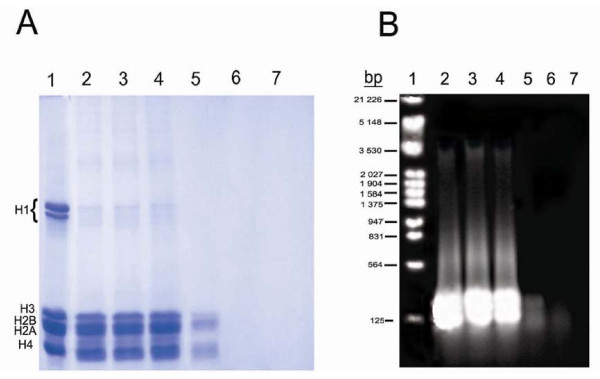
Figure 3.
A: 15% SDS gel electrophoresis of the supernatants obtained from the interaction of various concentrations of mitoxantrone with SE-chromatin. Lanes 2–7 are 0, 10, 20, 30, 40 and 60 μM of mitoxantrone respectively. Lane 1, calf thymus histones as a standard. B: Agarose gel of DNA extracted from the supernatants. Lane 1, EcoR1 DNA marker. Lanes 2–7 are the same as in gel A. Number of experiments was 3.

