
Figure 4.
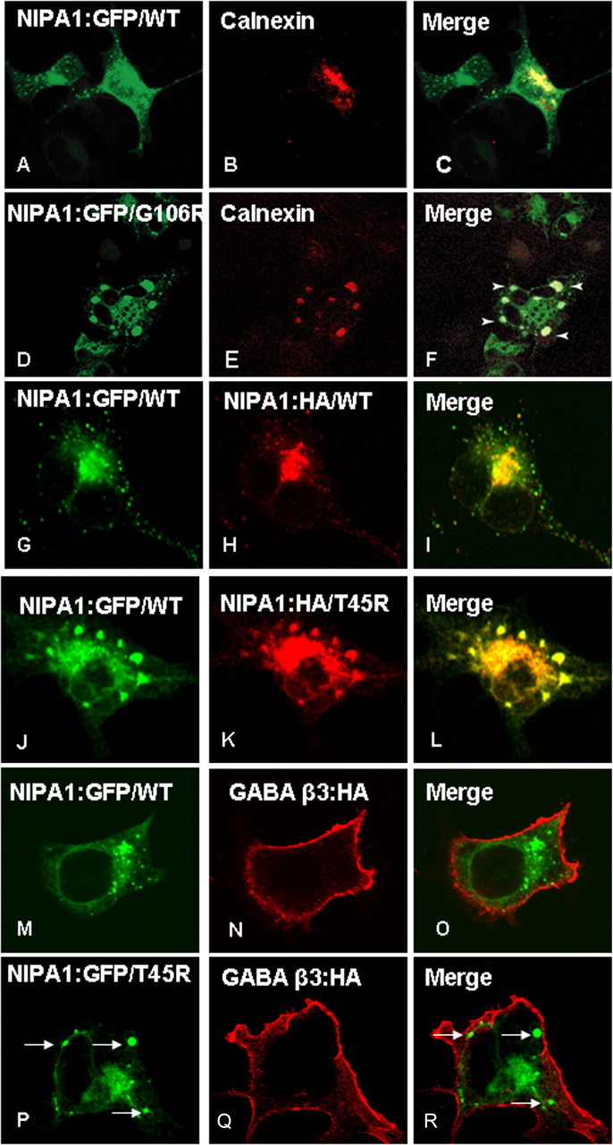
Endoplasmatic reticulum accumulation of mutant NIPA1. Overexpression of WT NIPA1::EGFP (A) shows a diffuse presence in the ER, which was visualized by ER marker anti-calnexin antibodies (B; C, overlay). Expression of a mutant NIPA1::EGFP (G106R mutation shown, D) resulted in the accumulation in ER, anti-calnexin antibodies (E; F, overlay) (arrowheads indicate NIPA1+ aggregates). The ER was abnormal with the presence of cystic structures. Overexpression in a “heterozygous state” showed that both WT and mutant forms of protein colocalized in ER. G–I show a control experiment with heterozygous expression of NIPA1::EGFP (G) and NIPA1:HA (H; I, merge), which did not show any effect of expressing both forms of the tagged protein on its localization. J–L show the expression of a mixture of WT and T45R, resulting in an alteration of WT NIPA1 subcellular localization, including sequestration in ER aggregates (J, WT NIPA1::EGFP; K, NIPA1T45R::HA; L, merge); G106R mutations had essentially an identical effect on WT NIPA1 protein (data not shown). The presence of mutant NIPA1 accumulation in ER (P–R, arrows indicate NIPA1 accumulation) did not affect the cell surface trafficking of the β-3 subunit of GABAA receptor; M–O shows the same GABA protein in the presence of WT NIPA1.