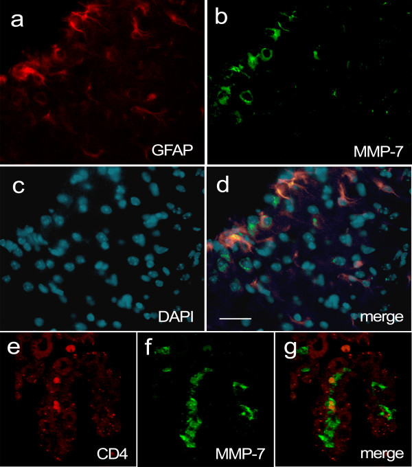
Figure 3.
MMP-7 immunoreactivity does not co-localize with the astrocyte marker GFAP, or CD4, in the CNS of wt mice during EAE. Brain sections were taken from a wt mouse 15 days after vaccination with MOG, when the EAE clinical score was 1. (a) MMP-7 (green) and (b) GFAP (red) immunopositive cells. (c) DAPI staining of nuclei. (d) Merged image showing that GFAP and MMP-7 do not co-localize to the same cells although there are interactions between these two immunopositive populations. Scale bar = 50 μm. (e) CD4 immunostaining in the choroid plexus. Intensity of the red signal was increased to show choroid plexus structures, from auto-fluorescence. (f) MMP-7 immunostaining (green) shows several immunopositive cells between endothelial sheets, on the vascular side. (g) Merged image of e and f shows that CD4+ and strong MMP-7 immunostaining do not co-localize. Arrow indicates continuity between an MMP-7 immunopositive cell and the lateral ventricle.

