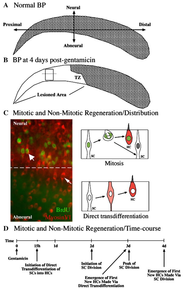
Figure 1. HC regeneration in the avian BP after short Gentamicin treatment.
A. HCs (stippling) are distributed throughout the undamaged BP. The neural side of the BP is indicated and overlies the cochlear ganglion. B. HCs are missing from the proximal BP at 4 days post-Gentamicin (no stippling). The zone of transition (TZ) between damaged and undamaged epithelium is indicated. C. Immunolabeling for MyosinVI (red) and BrdU (green) in the damaged BP, in the region shown in the rectangular box in B. The dotted line separates neural and abneural regions. BrdU-positive cells are densest in the neural (upper) region, although regenerated HCs (MyosinVI-positive) are distributed throughout the neural-abneural axis. HCs regenerated via mitosis have BrdU-labeled nuclei (thick arrow) and are densest in the neural side, while HCs regenerated via direct transdifferentiation have BrdU-negative nuclei (thin arrow) and are most common in the abneural side. The two mechanisms of HC regeneration are diagrammed on the right side of C. D. The time-course of mitotic and non-mitotic regeneration is outlined. d=days; h=hours.

