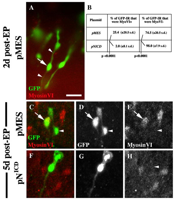
Figure 8. Overexpression of NICD prevents SCs from directly converting into HCs after damage.
This figure shows representative GFP-positive cells (green) in brightest point Z-projection images from BPs at 2 days (A) or 5 days (C–H) after transfection of negative control plasmid (pMES) or pNICD. Counterlabeling for MyosinVI is shown in red. A. GFP-positive cells at 2 days after transfection with pMES have SC-like features (long cytoplasmic processes [arrowheads] that are MyosinVI-negative; arrow points to nucleus). B. Quantitative data from cultures at 5 days post-transfection (C–H). C–E. GFP-positive cells at 5 days after transfection with pMES have either a HC-like morphology (MyosinVI-positive and fusiform or round) or a SC-like morphology. F–H. The vast majority of GFP-positive cells at 5 days after transfection with pNICD have a SC-like morphology. Images in C and F are shown in separate channels in D,E and G,H, respectively. Scale bar = 20 μm.

