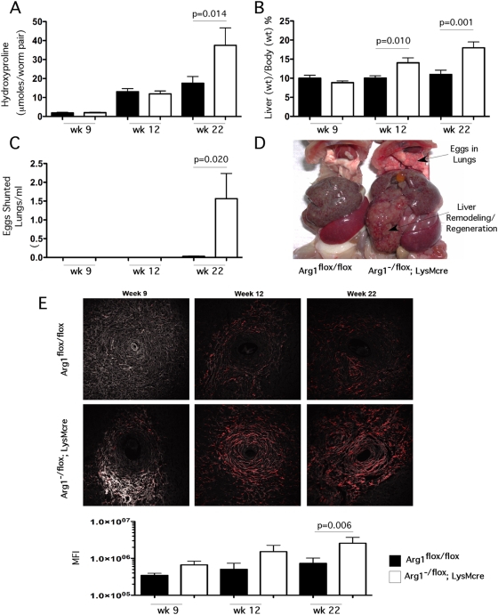
Figure 2. Arg1 expression is required to control fibrosis.
Control Arg1 flox/flox (filled bars) and Arg1 −/flox ;LysMcre (open bars) mice were exposed to 35 S. mansoni cercariae percutaneously. Mice (n = control/Arg1 −/flox ;LysMcre ) were sacrificed at weeks 9 (n = 14/14), 12 (n = 21/10), and 22 (n = 12/10) post-infection and analyzed for the development of fibrosis and portal hypertension. (A) Collagen was assessed by measuring hydroxyproline and normalizing to infectious worm pairs per mouse (Mean±SEM). (B) Total animal weight was compared with the weight of total excised liver to determine liver as a percent of body weight (Mean±SEM). (C) S. mansoni eggs within the lungs of control and Arg1 −/flox ;LysMcre mice as an measure of collateral vessel development and portal hypertension; eggs were enumerated by digesting lungs in 4% KOH at 37°C for 12 hours and 1ml of the suspension was counted in a Sedgwick-Rafter chamber (Mean±SEM). (D) Representative gross pathology of 22-week infected control and Arg1 −/flox ;LysMcre. Upper arrow indicates presence of S. mansoni eggs which were shunted into the lungs of Arg1 −/flox ;LysMcre mice. (E) Individual liver sections from Control Arg1 flox/flox (top) and Arg1 −/flox ;LysMcre (bottom) mice were analyzed for collagen via second harmonic emission (red). Representative granulomas from 9, 12, and 22 weeks post-infection with S. mansoni. All images were taken at 20× magnification. Mean fluorescence intensities for individual control (filled bars) and Arg1 −/flox ;LysMcre (open bars) mice at week 9 (n = 14/14), 12 (n = 21/10), and 22 (n = 12/9) weeks post-infection with S. mansoni. All assays were repeated three times with similar results.

