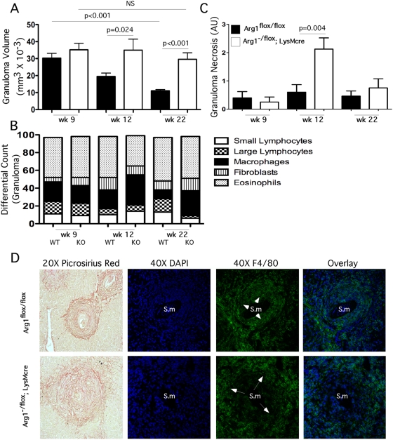
Figure 3. Macrophage-associated Arg1 inhibits granulomatous inflammation.
Week 9 (n = 14/14), 12 (n = 21/10), and 22 (n = 12/9) S. mansoni–infected control and Arg1 −/flox ;LysMcre mice were individually assessed for granuloma volume (Mean±SEM) (A), and individual populations of small/large lymphocytes, macrophages, fibroblasts, and eosinophils were enumerated (B). Individual granulomas from control (filled bars) and Arg1 −/flox ;LysMcre mice (open bars) were scored for granuloma-associated necrosis on a scale of 1–4 at weeks 9 (n = 14/14), 12 (n = 21/10) and 22 (n = 12/9) post-infection with S. mansoni (mean±SEM) (C). Representative granulomas from 12-week S. mansoni–infected control and Arg1 −/flox ;LysMcre mice were stained with Picrosirius Red 20× or DAPI (blue) and F4/80+ (green) and photographed at 40×. Arrows point to macrophage-rich regions. Sm = S. mansoni egg (D). All assays were repeated three times with similar results.

