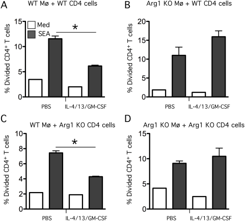
Figure 9. Arg1 is required to suppress schistosome-specific T cell proliferation.
2×10⁵ thioglycollate-elicited macrophages from (WT) control Arg1flox/flox (A,C) and (KO) Arg1 −/flox ;LysMcre mice (B,D) were pre-treated with a cocktail of IL-4/IL-13/GM-CSF at a concentration of 1 ng/ml of each cytokine or PBS. CD4+ cells were isolated from the livers of 9-week S. mansoni–infected WT (A,B) or Arg1 −/flox ;LysMcre mice (C,D), labeled with CFSE, and then added to the macrophage cultures at a concentration of 1×10⁵ cells per well in RPMI+10% FCS. Wells were either left unstimulated (Med) or stimulated with 20 µg/ml of SEA. After 84 hours, proliferation was analyzed by flow cytometry and represented as the percentage of CD4 cells that had divided in each of the conditions. The experiment was conducted twice with similar results.

